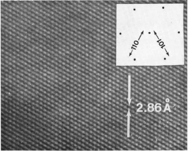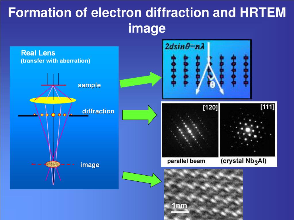

from a method called x-ray diffraction, applied typically to small samples of crystalline material. The spinel-based monoclinic model is also more accurate than the monoclinic nonspinel model. CRYSTALMAKER, CACHE EXCEL AT VISUALIZING MOLECULES. Thereby, not only the d-spacings but also the positions of the diffraction. Our work indicates that the traditional cubic spinel model is a more accurate model of γ-Al 2O 3 than the other models considered. Electron diffraction pattern from the area marked in c. The single-crystal SAED spot pattern showed symmetry consistent with both the cubic spinel and tetragonal nonspinel models, however, the Al cation distribution better matched the cubic spinel model based on the relative intensities of diffraction spots. were generated using CrystalMaker and SingleCrystalTM. d The selected are diffraction (SAED) pattern of it. The length and width of the electron transparent region of the lamella are approximately 5 m and 3 m, respectively. c A TEM overview of the lamella (show in Fig. (e) Selected area diffraction pattern obtained from the 001 zone. a, b Show the schmatic of imaging and diffraction patterns. (b) and (d) are diffraction simulation results from the 100 zone axis and (c) 110 zone axis.

Simulation results of electron diffraction using (b) P4/mmm, (c) P4/nbm, and (d) Cmmm crystal structure models 2-4. The lattice interplanar distances derived from the polycrystalline SAED pattern most closely matched the cubic spinel γ-Al 2O 3 model. data and qualitative trends in the electron-diffraction patterns reveal that the secondary X1a. Contrast-enhanced diffraction pattern is shown in the lower left. Single crystal and textured polycrystalline γ-Al 2O 3 thin films were synthesized by oxidation of NiAl(110) in air at 850 ☌ for 1 and 2 h, respectively, and used to evaluate the accuracy of two spinel-based and two nonspinel models by comparison of selected-area electron diffraction (SAED).


 0 kommentar(er)
